
Dr. Peter Derman
Texas Back Institute
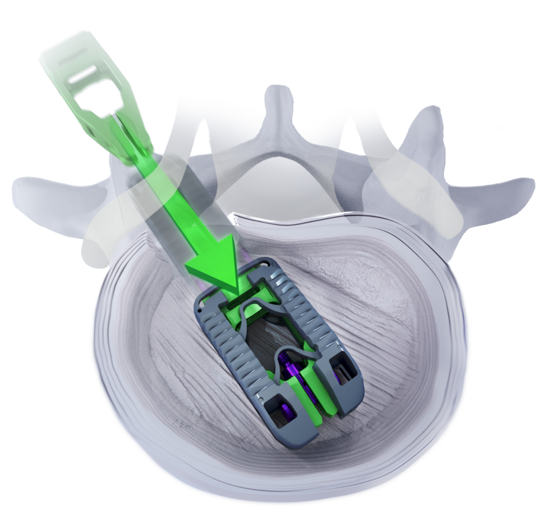
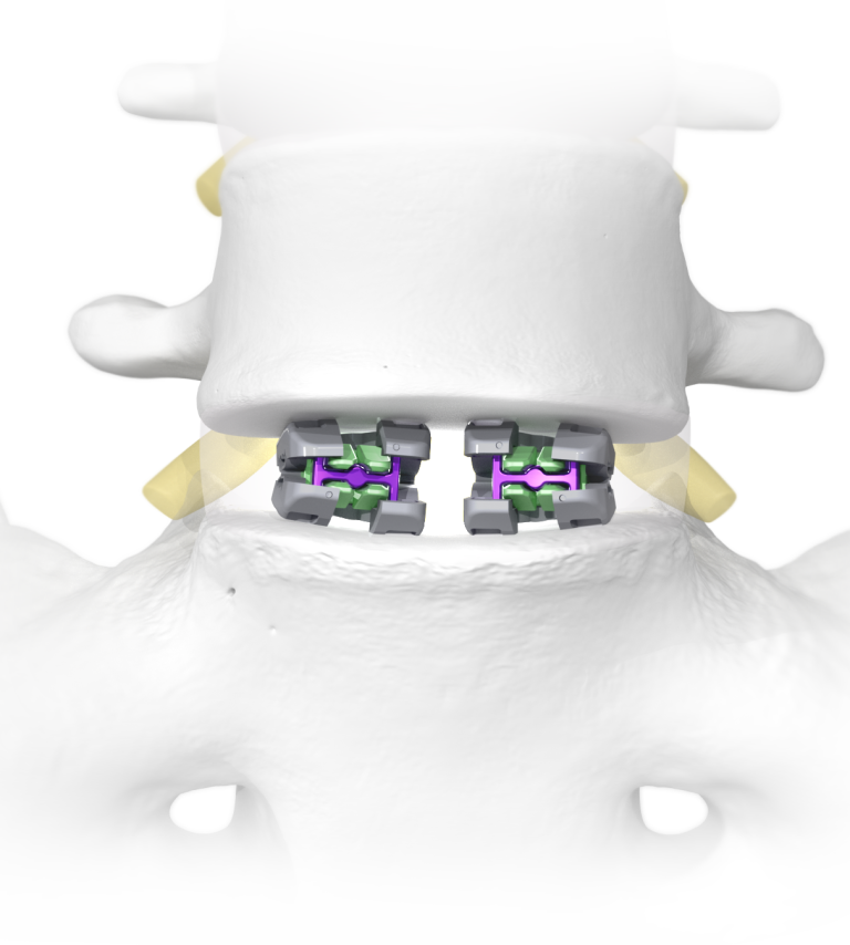
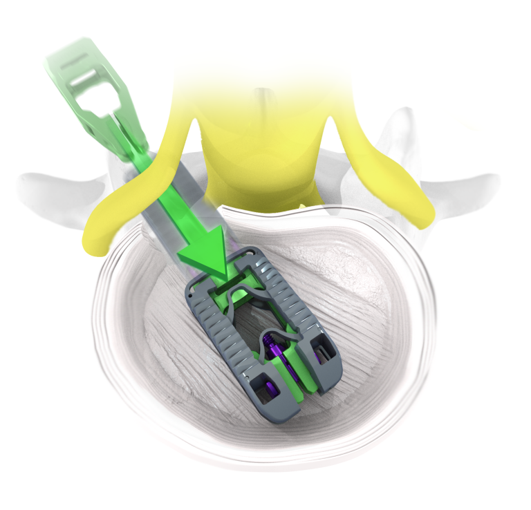
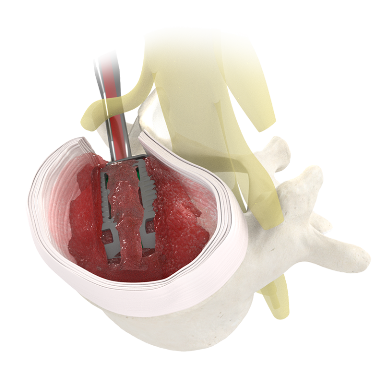
"These interbody cages feature multi-planar expansion, adaptive geometry, and open architecture designed to facilitate safer insertion and deployment - unique properties that can minimize subsidence and maximize fusion."
Dr. Stephen Tolhurst
Texas Back Institute
"With all titanium expandable cages, we can see subsidence due to point loading. TiHawk11 shines because of its Adaptive Geometry. The conformity of the TiHawk11 implant to the endplates when it is expanded allows for the patient's disc space to expand to its proper physiological height with the load better distributed across the entire surface of the implant. The end result is an expandable cage that conforms to the unique geometry of the disc space under load"

Dr. Kalman D. Blumberg
Florida Spine Specialists
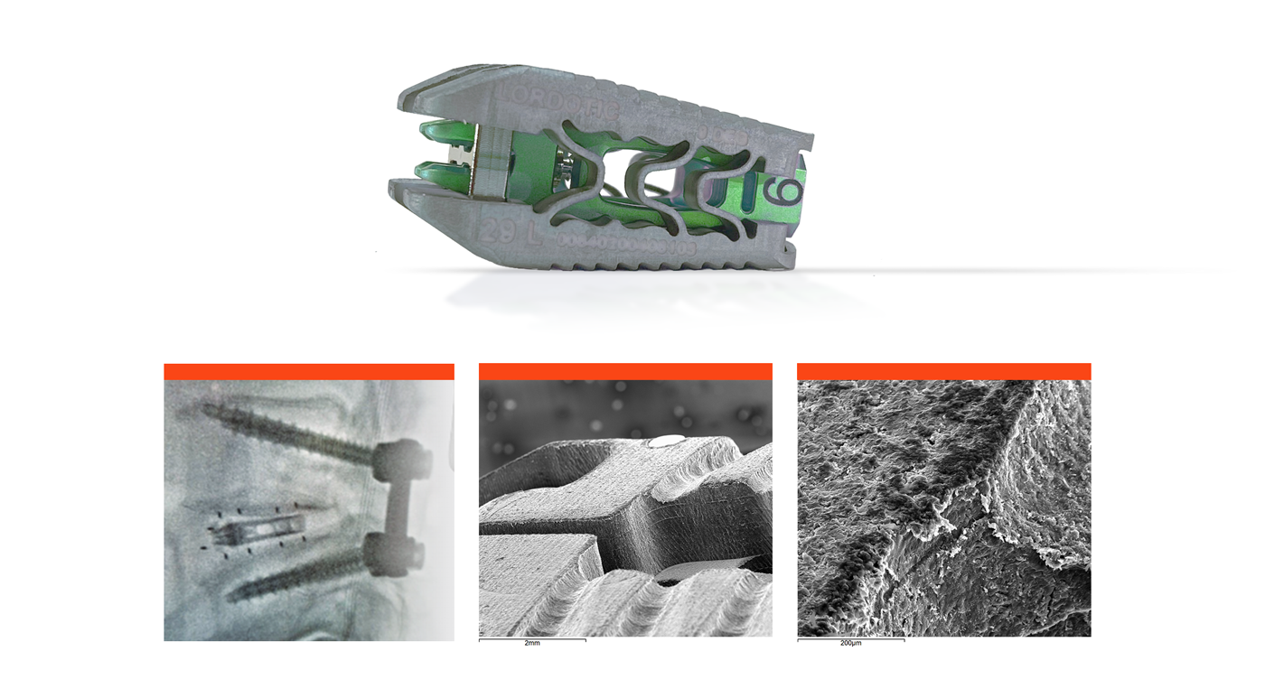
"The titanium-bonded TiHawk11 continues the solid performance of all the Accelus interbody devices produces to far. This latest version allows a minimal insertion profile compared to its expanded size, which is especially useful for my revision cases requiring a tight dissection. I have found the performance of the TiHawk11 to be unparalleled."